Nerves explained simply; It is a system of cables that delivery electrical current from the brain and spinal cord to the organs to provide movement and restore senses such as sensation, heat, pressure from the organs. Therefore, there are two types of nerves as motor function and sensory nerves.
The nerves in our fingers are pure sensory nerves. They are not related to movement. The purpose of repairing an injured finger nerve is to restore functional sensation and prevent the formation of painful cut nerve ending swelling called neuroma.
In order to understand nerve damage in patients presenting with finger nerve injuries, a neurological examination including evaluations such as sensation test and two point discrimination test should be performed before local anesthesia. Since the veins that provide the circulation to the fingers are located right next to the nerves, there is a high risk of injury. Therefore, veins and tendons that provide finger movement are also carefully examined. In order to avoid sequelae of delayed and missed nerve injury in children or patients who cannot be fully evaluated is to surgically evaluate the nerve and repair it if there is any injury.
Factors Positively Affecting the Treatment Results in Finger Nerve Injuries
- Early nerve injury repair
- Younger age patients
- Clean wound
- The nerve to be cut in a shape called as a guillotine incision.
- Non-smoking patients
The most important of these factors is age. While the treatment results are much better in children, there is no age limit for nerve repair. There is no time limit for repair of the sensory nerves in the early stages, but the earlier it is performed, the better the results. The retraction of the nerves is possible and adhesions in the surrounding tissues to make it difficult to bring the nerves end to end may occur after three weeks, which may adversely affect the results of the surgery.
Finger Nerve Repair with Microsurgery
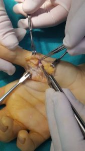
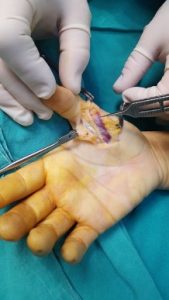
Microsurgical techniques are used in the repair of finger nerve injuries, which approximately have a diameter of 2 mm. With 10 times magnification under the microscope, suture materials with a thickness that can hardly be viewed with the naked eye and the nerves are sutured back together. If the cut ends of the nerves are too missing or retracted to come end to end, the nerve parts preferably taken from the leg are sutured as a bridge. Since the sutures applied after the surgery are very thin, a 3-week protective cast-splint is applied to protect the repair area.

